Brain Art: AAL Patchwork
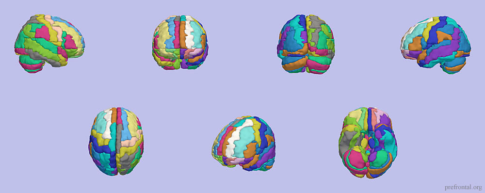
This is a rendering done in Slicer of the numerous brain regions defined in the Automated Anatomical Labeling (AAL) digital brain atlas. The AAL atlas was constructed through the identification of major and minor sulci on a T1 MRI with subsequent labeling based on anatomical location. It doesn’t have any relationship to cortical cytoarchitecture, but is still a useful tool to let you know what is in the neighborhood.
I first made this rendering several years ago and since that time I have really fallen in love with it. The picture really brings together the patchwork complexity of the cortical and cerebellar surface. This is an important point – that there is much more to the brain than just two halves or four lobes. It is also very clear that there are asymmetries between the two hemispheres of the brain. The shape of a region in the left hemisphere has little relationship with the configuration of its counterpart in the right hemisphere. I dig it.
If you click on the above image it will take you to a larger version you can download. Enjoy!
* Tzourio-Mazoyer N, Landeau B, Papathanassiou D, Crivello F, Etard O, Delcroix N, Mazoyer B, Joliot M. (2002). Automated anatomical labeling of activations in SPM using a macroscopic anatomical parcellation of the MNI MRI single-subject brain. Neuroimage 15(1): 273-289.
3 Responses to “Brain Art: AAL Patchwork”
Daniel Levy - April 20th, 2011
Your AAL rendering of the brain is quite graceful, and helps me understand the relationships of the AAL defined regions. Do you by any chance have a version in which the regions defined by colors are also labeled by name? It would be a great help.
Thanks
Unfortunately, I don’t have a labeled copy of the AAL rendering. The next time I am working with Slicer I will try to produce a higher-resolution image that I can annotate with the region labels. It would be a nice addition, and useful around our lab as well. ~Craig
Ying-hui Chou - May 31st, 2012
Your AAL rendering of the brain looks great.
Could I use one of your figures in my manuscript to show how the brain was parcelled?
If yes, how should I cite your work?
Thank you.
Adi Maron-Katz - May 5th, 2014
Hello,
Your picture is very nice.
may I use it in a poster for the coming OHBM conference?
Thanks in advance
Adi Maron-Katz
PhD student in Neuroscience